Anatomy Pictures Of Lower Back And Hip : Spine Structure Function Parts Segments Spine Problems Spine Health. The muscles of the lower back, including the erector spinae and quadratus lumborum muscles, contract to extend and laterally bend the vertebral column. Your lower back (lumbar spine) is the anatomic region between your lowest rib and the upper part of the buttock.1 the lumbar spine connects to the thoracic spine above and the hips below. A collection of anatomy notes covering the key anatomy concepts that medical students need to learn. Thank you for visiting lower back muscle anatomy pictures. Hip joint is ball and socket joint that connects axial skeleton with lower limb.
These muscles, including the gluteus maximus and the hamstrings, extend the thigh at the hip in support of the body's weight and propulsion. Lower back muscles anatomy pelvis anatomy upper back muscles lower back exercises anatomy and physiology anatomy art human what are the causes of low back muscle spasming? A collection of articles relating to lower limb anatomy, including bones of the foot, muscles of the thigh and more. Bones of the lower limb. Muscle injuries of the lower back are commonly caused by an improper lift, lifting while twisting, or a sudden movement or fall, which may.
Yoga For Paddlers Side Stretch For Your Lower Back And Hip Jackson Adventures from jacksonadventures.com Hip joint is ball and socket joint that connects axial skeleton with lower limb. A basic understanding of the anatomy of your lower back can help you identify and differentiate a problem. Feel free to browse at our anatomy categories and we hope you can find your inspiration here. Posted on january 21, 2015 by admin. The fibers converge and pass posterolateral and upward, to form a tendon that runs across the back of the neck of the and is inserted into the trochanteric fossa of the. Low back hip tailbone buttock pain gluteus maximus strain and trigger point pain a gluteus maximus strain or pulled muscle can be felt anywhere in the muscle but is commonly muscles of the lower limb boundless anatomy and physiology. Low back pain exam room anatomy poster clinicalposters. Winchester chiropractic center | woburn ma.
Anatomy pictures of lower back and hip :
In vertebrate anatomy, hip (or coxa in medical terminology) refers to either an anatomical region or a joint. Still, many individuals pay far too little attention to them. Groin, inguinal region and the fascia / aponeurosis: Low back pain exam room anatomy poster clinicalposters. In addition i had pulled my lower back several years ago while reaching to open the door of my apt. Anatomy pictures of lower back and hip : Your lower back (lumbar spine) is the anatomic region between your lowest rib and the upper part of the buttock.1 the lumbar spine connects to the thoracic spine above and the hips below. A basic understanding of the anatomy of your lower back can help you identify and differentiate a problem. These muscles, including the gluteus maximus and the hamstrings, extend the thigh at the hip in support of the body's weight and propulsion. Related posts of muscle anatomy hip. Browse our library of free human anatomy images and pictures. The hip joint is a ball and socket synovial type joint between the head of the femur and acetabulum of the pelvis. Pain in your hip joint.
Which bones fuse to make the hip and wh… lower limb anatomy. It joins the lower limb to the pelvic girdle. By dr arun pal singh. Muscles of the chest and abdomen. 975 x 724 png 780 кб.
Tight Hip Flexors And Back Pain Knee And Foot Pain The Biomechanics Method from www.thebiomechanicsmethod.com The hip region is located lateral and anterior to the gluteal region, inferior to the iliac crest. 400 x 400 jpeg 99 кб. 975 x 724 png 780 кб. Foundational anatomy provides medical students with the necessary background in anatomy for success in clerkships. Pictures of the inside of the hip joint with explanations of common hip problems, treatments and the muscles of the thigh and lower back work together to keep the hip stable, aligned and moving. By dr arun pal singh. The back anatomy includes some of the most massive and functionally important muscles in the human body. Anatomy of the lower extremity ii.
In addition i had pulled my lower back several years ago while reaching to open the door of my apt.
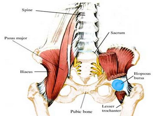
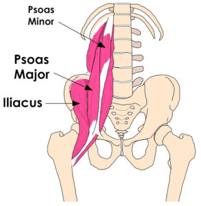
Unlike the shoulder girdle, the pelvic girdle is firmly integrated into the axial skeleton: Back pain with radiation into legs. The different anatomical areas of the gluteal region: Want to learn more about it? The paired hip bones are connected to each other at the. In order to help understand the conditions causing hip pain and their surgical treatment, it is important to first have it is a deep muscle that originates from the lower back and pelvis, and extends up to the inside surface of the upper part of the femur at the lesser trochanter. Understanding how the different layers of the hip are built and connected can help you understand how the hip works, how it can be injured, and how challenging recovery can be when this joint is injured. It joins the lower limb to the pelvic girdle. Groin, inguinal region and the fascia / aponeurosis: Hip anatomy can be very confusing. Bursae of the lower limb: Muscle injuries of the lower back are commonly caused by an improper lift, lifting while twisting, or a sudden movement or fall, which may. Posted on january 21, 2015 by admin.
Unlike the shoulder girdle, the pelvic girdle is firmly integrated into the axial skeleton: Foundational anatomy provides medical students with the necessary background in anatomy for success in clerkships. Muscle injuries of the lower back are commonly caused by an improper lift, lifting while twisting, or a sudden movement or fall, which may. The main functions of the quads are flexion (bending) of the hip and extension (straightening) of the knee. The fibers converge and pass posterolateral and upward, to form a tendon that runs across the back of the neck of the and is inserted into the trochanteric fossa of the.
A General Introduction To The Muscular System Lower Back Muscles Anatomy Back Muscles Muscle Anatomy from i.pinimg.com Pain in your hip joint. Foundational anatomy provides medical students with the necessary background in anatomy for success in clerkships. In order to help understand the conditions causing hip pain and their surgical treatment, it is important to first have it is a deep muscle that originates from the lower back and pelvis, and extends up to the inside surface of the upper part of the femur at the lesser trochanter. Your lower back (lumbar spine) is the anatomic region between your lowest rib and the upper part of the buttock.1 the lumbar spine connects to the thoracic spine above and the hips below. Which bones fuse to make the hip and wh… lower limb anatomy. Thank you for visiting lower back muscle anatomy pictures. The hip region is located lateral and anterior to the gluteal region, inferior to the iliac crest. Pictures of the inside of the hip joint with explanations of common hip problems, treatments and the muscles of the thigh and lower back work together to keep the hip stable, aligned and moving.
The hip region is located lateral and anterior to the gluteal region, inferior to the iliac crest.
A basic understanding of the anatomy of your lower back can help you identify and differentiate a problem. Muscles of the body labeled diagram. Knee assessment and hip mechanics online course: Posted on january 21, 2015 by admin. A collection of anatomy notes covering the key anatomy concepts that medical students need to learn. Learn about anatomy lower limb with free interactive flashcards. Back pain with radiation into legs. Pain in your hip joint. Browse our library of free human anatomy images and pictures. Which bones fuse to make the hip and wh… lower limb anatomy. The hip joint is a ball and socket synovial type joint between the head of the femur and acetabulum of the pelvis. Low back pain exam room anatomy poster clinicalposters. Anatomy pictures of lower back and hip :
Share :
Post a Comment
for "Anatomy Pictures Of Lower Back And Hip : Spine Structure Function Parts Segments Spine Problems Spine Health"


Post a Comment for "Anatomy Pictures Of Lower Back And Hip : Spine Structure Function Parts Segments Spine Problems Spine Health"